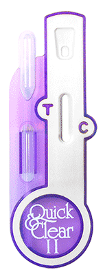
Storage and Stability
Store at room temperature (15° to 30°C / 59° to 86°F). The test device must remain in the foil pouch until ready for use. Sealed pouches are good until the expiration date listed on the pouch.
Precautions
1. Test devices should remain in the sealed pouch until ready for use.
2. All specimens should be considered potentially hazardous and handled in the same
manner as an infectious agent.
3. The test device should be discarded in a proper biohazard container after testing.
4. Do not use test kit beyond the expiration date.
Specimen Collection and Preparation
Urine specimens should be collected in a clean, dry container. Randomly collected
specimens can be used. However, the first morning urine generally contains the highest
concentration of hCG. If urine samples exhibit visible precipitates, the specimen should be
filtered, centrifuged, or allowed to settle to obtain clear aliquots.
Specimen Storage
Specimens may be refrigerated (2° -
8°C) and stored up to 72 hours prior to assay. If specimens are refrigerated, they must be brought
to room temperature before testing. If testing is delayed more the 48 hours, the specimens should
be frozen. Frozen specimens should be thawed and mixed before testing.
Directions For Use
View proper drop placement/timing here
Allow specimen and/or controls to reach room temperature
(15°C to 30°C) prior to testing.
1. Remove the test device from the sealed pouch and place it on a level, dry surface.
2. Dispense 3 drops (approx. 0.12ml) of the urine specimen into the sample well. Wait for
colored lines to appear.
3. Read results after 3 minutes. Some positive results may be observed in 60 seconds or less
depending on the concentration of hCG. Specimens containing levels of hCG below 20mIU/ml may show
color development over time, therefore, do no read results after 10 minutes.
Interpretation of Results
Negative Results
The test is negative if only one colored line appears in the Control Zone. This indicates the
absence of detectable levels of hCG.
Positive Results
The test is positive if two colored lines appear. One colored line will appear in the Test Zone and
one in the Control Zone. This indicates the specimen contains detectable levels of hCG. Any
colored line in the Test Zone should be considered positive. Specimens containing levels of hCG
below 20 mIU/ml may show color development over time. The colored line in the Control Zone may be
lighter or darker in color than the line in the Test Zone.
Invalid Results
The test is invalid if a colored line fails to appear in the Control Zone, even if a colored line
appears in the Test Zone. If this occurs, add 2 additional drops of specimen and wait 3 minutes. If
a colored line still does not appear in the Control Zone, the test is invalid and should be
repeated with a new test device.
Limitations
1. A number of conditions other than pregnancy may
produce elevated levels of hCG such as trophoblastic disease, choriocarcinoma, embryonal cell
carcinoma, Islet cell tumors, and other carcinomas.4
2. If urine specimens are too dilute, false negative results
may occur due to low levels of hCG below the sensitivity of the test. If pregnancy is still
suspected, a first morning urine should be collected 48 hours later and tested.
3. False positive results may be due to detectable levels
of hCG remaining several weeks after a normal pregnancy, delivery by cesarian section, spontaneous
or therapeutic terminations.5
4. Very low levels of hCG may be found in ectopic
pregnancies6 and additional testing may be necessary using a quantitative assay.
5. Natural termination occurs in 22% of clinically
unrecognizable pregnancies, and 31% of pregnancies overall.7 This may produce positive results when
testing early in the pregnancy followed by negative results after the natural termination. In
cases exhibiting weak positive results, it is good laboratory practice to confirm results by
retesting with a first morning urine 48 hours later.
6. All clinical and laboratory findings should be evaluated
before making a definitive diagnosis.
REFERENCES
1. Braunstein GD, Rasor J, Adler D, Danzer H, Wade ME:
Serum human chorionic gonadotropin levels through normal pregnancy. Am J Obstet Gynecol, 126:
678-681,
1976.
2. Schwartz S, Berger P, and Wick G: Epitope-selective
monoclonal antibody based immunoradiometric assay of predictable specificity for differential
measurement of choriogonadotropin and its subunits, Clin Chem 31:1322-
1328, 1985.
3. Kaplin LA, Pesce AJ. Clinical Chemistry Theory, Analysis,
& Correlation, 2nd Edition, Missouri, 1989, C.V. Mosby
Co., p. 944.
4. Jacobs DS, et al: Laboratory Test Handbook, 2nd Edition,
Ohio, Lexi-Comp Inc., pp. 224, 305-307, 1990.
5. Steier JA, Bergso p, Myking OL: Human chorionic
gonadotropin in maternal plasma after induced abort., spontaneous abortion, and removed ectopic
pregnancy. Obstet Gynecol 64: 391-394, 1984.
6. Thorneycroft IH: When you suspect ectopic pregnancy.
Diagnosis January: 67-82, 1976.
7. Wilcox EG, Weinberg CR, O'Connor JF, et al: Incidence of
early loss of pregnancy. N Eng J Med 319: 189-194,
1988.
8. Chard T, Pregnancy Tests: A Review. Human
Reproduction, 7:701-710, 1992.